
[Medically reviewed by Dr. Garvit Chitkara, MBBS, DNB (Gen Surg), HBNI Fellowship in Breast Oncology (TMH), Clinical Observership in Breast Oncoplasty at Memorial Sloan Kettering Cancer Center – Consultant Breast Surgical Oncologist, Mumbai, with 17+ years of experience. ]
If your mammogram report mentions “breast calcifications,” it is natural to feel uneasy. In most cases these are small deposits of calcium salts in the breast tissue, and they are among the most common findings on a mammogram. The large majority are benign — in reported series, roughly 80% of calcifications seen on mammography reflect harmless, non-cancerous processes. A minority of patterns, however, do warrant a closer look, which is why radiologists describe them so carefully.
Dr. Garvit Chitkara, a breast cancer surgeon in Mumbai, sees this question come up often: patients are rarely sure whether a calcification result means something is wrong. Here is what these deposits are, how they are read on a mammogram, what the BI-RADS number on your report actually means, and when a biopsy is advised.
“Most breast calcifications seen on a mammogram turn out to be benign, and simply reflect normal changes in breast tissue over time. That said, a few specific patterns do call for a closer diagnostic work-up, which is why every mammogram should be reviewed carefully by a qualified breast specialist.” — Dr. Garvit Chitkara, Breast Cancer Surgeon in Mumbai
Not sure what your report actually means for you? Get it reviewed by a specialist who can explain your report in plain terms.
Key Takeaways
- Breast calcifications are calcium deposits in breast tissue. They are a common mammographic finding and are not a disease in themselves.
- Around 80% of calcifications seen on mammography are benign.
- They are almost never felt as a lump and do not cause pain. They are picked up on imaging.
- What matters is not the calcium itself but the shape (morphology) and arrangement (distribution) of the deposits.
- The BI-RADS category on your report translates directly into a recommended next step: routine screening, short-interval follow-up, or biopsy.
- Even among calcifications suspicious enough to biopsy, roughly 4 out of 5 turn out to be benign.
- Calcifications have nothing to do with how much calcium you eat or with calcium supplements.
What Are Breast Calcifications?
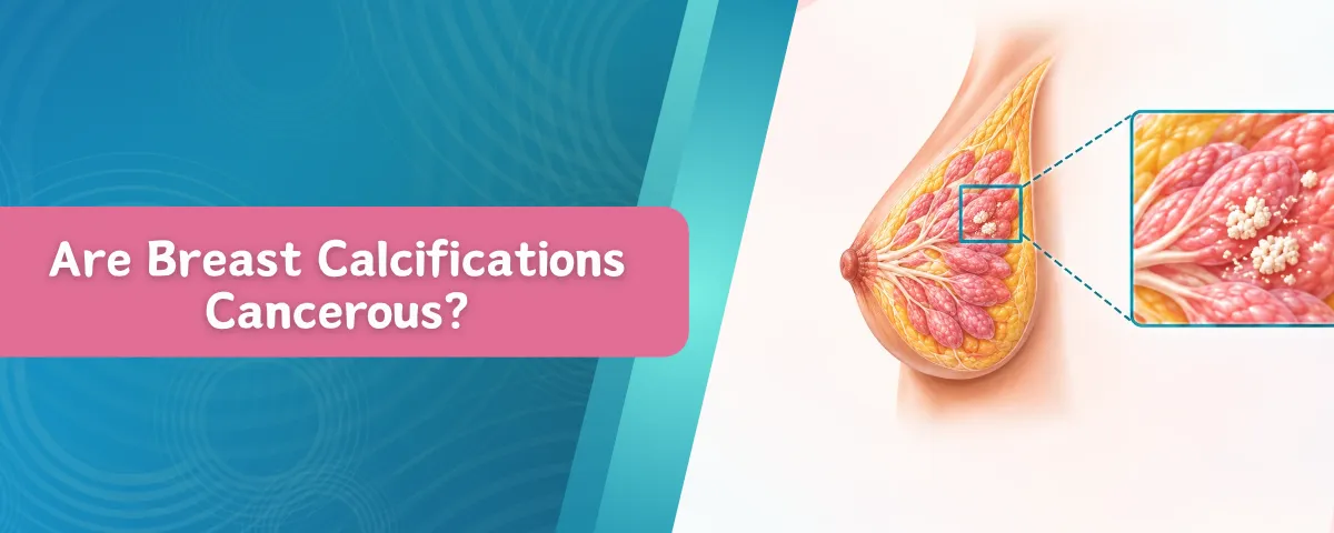
Breast calcifications are tiny deposits of calcium salts that develop within breast tissue. They show up as bright white specks or flecks on a mammogram because calcium absorbs X-rays strongly. They become more common with age and are found most often in women over 50, usually as an incidental finding during a routine mammogram.
Most women with breast calcifications have no symptoms at all. The deposits are far too small to feel, so they are not a cause of breast lumps and they do not cause pain. They are almost always invisible on clinical examination and are detected only on imaging.
Breast calcifications are not a disease in their own right. They are a sign of an underlying process, and in the overwhelming majority of cases that process is entirely benign. The important question is never “are there calcifications?” but “what do they look like, and how are they arranged?”

What Causes Breast Calcifications?
Calcium deposits in the breast can form for many reasons, almost all of them harmless:
- Normal ageing of breast tissue and involution of the milk-producing glands
- Benign conditions such as fibrocystic change, cysts and fibroadenomas — an old fibroadenoma often develops coarse, popcorn-like calcification
- Previous breast surgery, biopsy, radiotherapy or injury, which can leave dystrophic calcification or scar-related change
- Fat necrosis following trauma to the breast
- Previous infection or inflammation, such as mastitis, and duct ectasia
- Calcium deposits in the walls of small breast arteries (vascular calcification), a change that mirrors arterial calcification elsewhere in the body
- Calcified secretions inside the milk ducts
- Skin deposits, including those in sweat glands, and even talcum powder or deodorant residue on the skin, which can mimic calcification on the image
- Less commonly, cell turnover and debris within the milk ducts associated with ductal carcinoma in situ (DCIS)
One thing that does not cause them: dietary calcium. Calcium in your diet, calcium supplements and vitamin D have no bearing on whether breast calcifications form. This is one of the most common misconceptions patients bring to the clinic.
Macrocalcifications vs Microcalcifications: What’s the Difference?
Traditionally, radiologists divided calcifications by size. Macrocalcifications are larger, coarser deposits, generally more than 0.5 mm across, that are easy to see on a standard mammogram. They are almost always linked to ordinary changes such as ageing tissue, an old fibroadenoma, a previous injury or past surgery. They very rarely need anything beyond routine screening.
Microcalcifications are much smaller deposits, generally under 0.5 mm, which may appear scattered or in groups. Most of these are benign too. But because early changes in the milk ducts can produce them, their shape, size, number and arrangement are assessed closely, and a minority prompt further imaging or a biopsy.
It is worth knowing that current practice has moved beyond a simple size split. The fifth edition of the ACR BI-RADS atlas groups calcification descriptors into just two categories — typically benign and suspicious — based on morphology and distribution rather than size alone. So a radiologist reporting your mammogram today is looking less at “big versus small” and more at “what shape are they, and how are they laid out?”

Which Calcification Patterns Are Benign, and Which Are Suspicious?
This is the part of the report that actually drives the decision. The ACR BI-RADS lexicon lists specific descriptors on each side:
| Typically benign calcifications | Suspicious calcifications |
| Skin (dermal) and vascular calcifications | Amorphous (hazy, without a clear shape) |
| Coarse or popcorn-like, as seen in a degenerating fibroadenoma | Coarse heterogeneous |
| Large rod-like (secretory) calcifications | Fine pleomorphic (varied shapes and sizes) |
| Round and punctate calcifications | Fine linear or fine linear branching |
| Rim (eggshell) and lucent-centred calcifications | Distribution matters too: linear and segmental patterns are the most concerning, followed by grouped and regional |
| Dystrophic calcifications after surgery, radiotherapy or injury |
|
| Milk of calcium within tiny cysts |
|
| Suture calcifications |
|
Distribution is described as diffuse, regional, grouped, linear or segmental. Diffuse calcifications scattered throughout both breasts are almost always benign. “Grouped” (historically called “clustered”) is used when at least five calcifications sit within about 1 cm of each other. Linear and segmental distributions follow the path of a duct or a duct system, which is why they raise more concern.
Change over time matters as well. Calcifications that are new, increasing in number, or extending along a duct since your last mammogram are taken more seriously than identical findings that have been stable for years — which is why bringing your previous mammogram films or reports to every appointment genuinely changes the interpretation.
Understanding BI-RADS Categories for Calcifications
BI-RADS — the Breast Imaging Reporting and Data System, developed by the American College of Radiology — is a standard vocabulary that lets every radiologist report a mammogram the same way. Each examination ends with a single category from 0 to 6, and each category carries a defined likelihood of cancer and a recommended next step.
| BI-RADS | What it means | Likelihood of cancer | Usual next step |
| 0 | Incomplete assessment | Not yet assigned | Additional views, magnification or ultrasound, or comparison with older films |
| 1 | Negative | Essentially 0% | Routine screening |
| 2 | Benign | Essentially 0% | Routine screening |
| 3 | Probably benign | 2% or less | Short-interval follow-up, usually at 6 months, then periodic surveillance for 2–3 years |
| 4 | Suspicious | More than 2% to under 95% | Tissue sampling (biopsy). Often reported as 4A (>2–10%), 4B (>10–50%) or 4C (>50–<95%) |
| 5 | Highly suggestive of malignancy | 95% or higher | Biopsy, with treatment planning to follow |
| 6 | Known, biopsy-proven cancer | Confirmed | Imaging to guide treatment already under way |
A BI-RADS number is not a diagnosis. It is a statement of probability and a management recommendation: watch, re-scan, or sample. Two points are worth underlining. First, BI-RADS 0 simply means the radiologist needs more pictures before deciding — it is not a warning sign. Second, BI-RADS 4 spans an enormous range, from just over 2% at the low end to nearly 95% at the top, which is exactly why the 4A, 4B and 4C subdivisions exist and why you should ask which one applies to you.

Are Breast Calcifications a Sign of Cancer?
Usually not. Most calcifications are benign and reflect natural changes in breast tissue over time. But the relationship runs the other way round in an important sense: while few calcifications turn out to be cancer, calcifications are one of the most valuable early signs of cancer that mammography can detect.
The figures are worth knowing:
- Around 80% of ductal carcinoma in situ (DCIS) cases show up on mammography as calcifications, and approximately 96% of DCIS diagnosed on mammography is found through biopsy of calcifications.
- DCIS accounts for roughly 20–25% of breast cancers detected at screening mammography.
- Calcifications are the only visible sign in a substantial share of breast cancers that cannot be felt — reported at around 41–55% of non-palpable malignancies.
- Working in the opposite direction: when a suspicious group of calcifications is biopsied, the positive predictive value is roughly 20%. Around four out of five such biopsies come back benign.
So a calcification finding that leads to a biopsy is far more likely to end in reassurance than in a cancer diagnosis. New, tightly grouped, irregular, or linear and branching microcalcifications are the ones that need evaluation, precisely because they can be the first and only sign of an early, highly treatable change such as DCIS. Calcifications reported as BI-RADS 4 or 5 therefore need tissue sampling, because imaging alone cannot reliably separate benign from malignant in that range.
Wondering where your own report fits on this scale? A short conversation can settle it either way.
How Are Suspicious Calcifications Diagnosed?
When calcifications fall into BI-RADS 0, 4 or 5, a few structured steps follow.
Magnification views
Spot magnification mammography, usually in craniocaudal and lateral projections, enlarges the area so the shape, number and spread of each deposit can be judged properly. A true lateral view also helps identify milk of calcium, which characteristically layers out and is entirely benign. Quite a number of findings are downgraded to benign at this stage, with no biopsy needed.
Stereotactic or tomosynthesis-guided biopsy
Calcifications usually cannot be seen on ultrasound, so they are sampled under mammographic guidance (stereotactic or tomosynthesis-guided) rather than ultrasound guidance.
A vacuum-assisted breast biopsy is the preferred technique for calcifications: it retrieves a larger volume of tissue through a single small skin nick, under local anaesthesia, with minimal discomfort and no stitches. It samples calcifications more reliably than a standard core needle biopsy and reduces the chance that the result has to be revised after surgery.
Specimen radiography and marker clip
The tissue removed is X-rayed on the spot to confirm that the calcifications are actually present in the sample — without this step, a benign result cannot be trusted. A tiny titanium marker clip is then placed at the biopsy site so the area can be found again on future mammograms, or localised before surgery if that becomes necessary. The clip is safe, permanent and compatible with MRI.
Radiology–pathology correlation
Finally, the pathology result is checked against what the imaging showed. If a mammogram looked strongly suspicious but the pathology came back bland, the result is called discordant, and further sampling or surgical excision is advised rather than reassurance.

What Happens After the Biopsy?
Pathology results fall into three broad groups, and the third is the one most articles leave out.
- Benign. The great majority. Fibrocystic change, a fibroadenoma, fat necrosis, duct ectasia or simple calcified secretions. You return to routine screening, often with one confirmatory mammogram first.
- High-risk or borderline lesions. Atypical ductal hyperplasia, flat epithelial atypia, lobular neoplasia and similar findings. These are not cancer, but a proportion are upgraded to DCIS or invasive cancer when the area is fully excised, so surgical excision or close surveillance is usually recommended, and the finding also carries implications for your future screening.
- DCIS or invasive cancer. Treatment is then planned individually with a breast surgical oncologist — most often breast-conserving surgery, with decisions about radiotherapy, sentinel node assessment and hormone therapy based on the final pathology. DCIS in particular carries an excellent prognosis when treated.
If the result is benign, no treatment is needed. Calcifications are not removed simply for being there, and they do not need to be “cleared.” If the result points to a benign breast condition, management is directed at that condition, not at the calcium.
When Should You Consult a Breast Specialist?
A few situations are worth taking to a specialist rather than filing away:
- Your report mentions BI-RADS 0, 3, 4 or 5
- A follow-up mammogram or additional views have been advised
- A biopsy has been suggested, or you want a second opinion before agreeing to one
- The calcifications are described as new, increased, grouped, linear, segmental, pleomorphic or amorphous
- You have a family history of breast or ovarian cancer, a known BRCA1/BRCA2 mutation, or previous chest radiotherapy
- You have a lump, skin change or nipple discharge alongside the calcification finding
- You simply do not understand what the report says — a reasonable and common reason to come in
A calcification result by itself is rarely urgent. It is, however, worth understanding rather than setting aside, and worth acting on within weeks rather than months if follow-up has been recommended.
Don’t let an unanswered line in your report sit around longer than it needs to.
FAQs
Q1. Are breast calcifications always benign?
Q2. Do breast calcifications cause pain or a lump?
Q3. Is a biopsy always required for suspicious calcifications?
Q4. Can breast calcifications turn into cancer over time?
Q5. Does eating calcium or taking supplements cause breast calcifications?
Q6. Do breast calcifications go away on their own?
Q7. Can calcifications be seen on ultrasound or MRI instead of a mammogram?
Q8. I had a benign biopsy for calcifications. What happens next?
Q9. Should calcifications be removed?
References
- American College of Radiology. ACR BI-RADS Atlas, Breast Imaging Reporting and Data System, 5th edition. Reston, VA: American College of Radiology; 2013.
- Rao AA, Feneis J, Lalonde C, Ojeda-Fournier H. A pictorial review of changes in the BI-RADS fifth edition. RadioGraphics. 2016;36(3):623–639.
- Berg WA, et al. Outcomes of BI-RADS category 3 lesions in the National Mammography Database. Radiology. 2020.
- Grimm LJ, et al. Growth dynamics of mammographic calcifications: differentiating ductal carcinoma in situ from benign breast disease. Radiology. 2019;292(1):77–83.
- Burnside ES, et al. Use of microcalcification descriptors in BI-RADS 4th edition to stratify risk of malignancy. Radiology. 2007;242(2):388–395.
- Virnig BA, et al. Epidemiology of ductal carcinoma in situ. Ann Transl Med / NIH review, 2016.
- Hoffman DI, et al. Noncalcified ductal carcinoma in situ: rate and predictors of upgrade to invasive carcinoma. Academic Radiology. 2021.
- National Comprehensive Cancer Network (NCCN). Clinical Practice Guidelines in Oncology: Breast Cancer Screening and Diagnosis. Current version.
- Radiological Society of North America / American College of Radiology. RadiologyInfo.org — Mammography and breast calcifications: patient information.
About the Author
Dr. Garvit Chitkara is a Consultant Breast Surgical Oncologist practising in Mumbai, with a focus on breast cancer surgery, oncoplastic and breast-conserving procedures, and the diagnosis and management of benign breast disease, including the evaluation of mammographic calcifications and image-guided breast biopsy.
Dr Garvit Chitkara has 17+ years of experience as a doctor and has been practicing Breast Surgical oncology for a decade. After getting trained in breast oncology from Tata Memorial hospital he practiced as an Associate Director at TMH and has also trained many students.
Disclaimer: The information shared in this content is for educational purposes only and is not a substitute for individual medical advice. Please consult a qualified breast specialist about your own mammogram report.