
Discovering a breast lump while breastfeeding can understandably cause concern. Breastfeeding and the hormonal changes around pregnancy can alter the breast tissue and may cause several benign conditions, including engorgement, inflammatory breast changes, galactocele, fibroadenoma, and lactating adenoma.
Most breast lumps during lactation are not cancer. However, a new, persistent or enlarging lump should not be assumed to be a breastfeeding-related change. Breast cancer can occur during lactation, and clinical examination alone may not reliably distinguish a benign condition from malignancy.
If a lump persists, increases in size, or is associated with other concerning changes, it should be assessed by a qualified clinician. Imaging should not be unnecessarily delayed simply because a woman is breastfeeding.
“Most breast lumps during breastfeeding are caused by benign conditions. However, a new, persistent or enlarging lump should not be ignored and should be assessed by a qualified breast specialist.” — Dr. Garvit Chitkara, Breast Cancer Surgeon in Mumbai
If a breast lump doesn’t go away after feeding or expressing milk, don’t ignore it. Get it checked by a doctor for timely care.
Common Causes of Breast Lumps During Breastfeeding
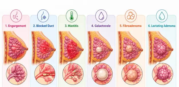
A breast lump during breastfeeding can have several causes. Identifying the cause is important because management differs depending on the underlying condition.
- Engorgement: When the breasts become overly full, they may feel firm, swollen, heavy, or lumpy. This usually improves as feeding patterns settle.
- Inflammatory breast changes / a “plugged” or “blocked” duct: Localised inflammation and narrowing of milk ducts can cause a tender, firm area. The older term “blocked duct” is commonly used by patients, but breast inflammation is more complex than simply a duct becoming blocked.
- Mastitis: Mastitis can involve inflammation of the breast and, in some cases, bacterial infection. It may cause a painful area, redness, swelling, warmth, and systemic symptoms such as fever or chills. A breast abscess can develop in some cases and requires medical assessment.
- Galactocele: A galactocele is a benign milk-filled cyst that can occur during lactation, often related to ductal obstruction.
- Fibroadenoma: A benign breast mass that may become more noticeable or enlarge during pregnancy and lactation because of hormonal influences.
- Lactating adenoma: A benign, hormonally influenced breast lesion that can present as a rapidly growing, well-circumscribed mass during pregnancy or lactation. Because it can resemble other breast masses, imaging and sometimes biopsy may be required.
If you notice a new lump, particularly one that persists or enlarges, do not rely on how it feels alone to determine whether it is benign. A clinical assessment is the safest way to decide whether imaging or further investigation is needed.
What Are the Symptoms of a Breast Lump During Breastfeeding?
A lump may feel different depending on its cause. It can be soft, firm or hard and may or may not be painful.
- A soft, firm or hard lump
- Pain or tenderness
- Breast swelling or fullness
- Localised redness or warmth
- Fever, chills or feeling unwell, particularly when mastitis is suspected
- A lump that persists or increases in size
- Skin dimpling, thickening or other visible changes
- New nipple inversion or persistent, unusual nipple discharge
Symptoms alone cannot reliably tell whether a lump is benign or malignant. If a palpable lump does not settle, is enlarging, or is clinically concerning, seek medical evaluation.
How Is a Breast Lump Evaluated During Breastfeeding?
Breastfeeding does not prevent a woman from having appropriate breast imaging. Ultrasound is commonly used as an initial imaging test for a palpable breast mass during lactation, particularly in younger women. Depending on age and clinical findings, diagnostic mammography or digital breast tomosynthesis may also be appropriate.
If imaging shows a suspicious finding, additional imaging or a biopsy may be recommended. These decisions are made according to the individual’s age, examination findings, imaging appearance and clinical risk.
Women should not stop breastfeeding solely because they need breast imaging. The appropriate investigation should be discussed with the treating clinician and radiologist.
How Can You Reduce the Risk of Breastfeeding-Related Breast Problems?
Not every breast lump can be prevented, but supportive breastfeeding practices may help reduce some common problems.
● Feed responsively according to the baby’s needs rather than following a rigid schedule.
● Pay attention to comfortable positioning and a good latch; seek lactation support if feeding is painful or difficult.
● Avoid unnecessary or excessive pumping or attempts to repeatedly “empty” the breast, as excessive breast stimulation may contribute to oversupply and inflammation.
● Avoid sustained pressure on the breast, including very tight bras or clothing.
● Maintain normal hydration and a balanced diet; extra fluids are not a specific treatment for breast lumps.
● If you develop symptoms of mastitis or a persistent lump, seek medical advice rather than relying on repeated breast massage or aggressive milk removal.
When Should You See a Doctor for a Breast Lump?

You should seek medical advice if you notice a new breast lump that is persistent, enlarging or otherwise concerning. Do not wait for a fixed period such as one or two weeks if the lump is clinically suspicious.
- A new lump that persists or increases in size
- A firm or fixed lump or a lump that feels different from the surrounding breast tissue
- Persistent redness, warmth, swelling or significant pain
- Fever, chills or feeling unwell, particularly with breast redness or tenderness
- Bloody, spontaneous, unilateral or persistent nipple discharge
- Skin dimpling, thickening or other new skin changes
- New nipple inversion or another persistent change in the nipple
Prompt assessment is particularly important when a lump does not behave like a temporary breastfeeding-related change or when symptoms are worsening.
FAQs
Q1. How long should a breast lump last during breastfeeding?
Some breastfeeding-related changes, such as engorgement or inflammatory symptoms, may improve over several days. However, there is no safe universal waiting period for a palpable lump. If a new lump persists, enlarges or is concerning on examination, it should be evaluated rather than waiting for one to two weeks.
Q2. Is every breast lump during breastfeeding a sign of breast cancer?
No. Many breast lumps during lactation are caused by benign conditions such as engorgement, inflammatory breast changes, galactocele, fibroadenoma or lactating adenoma. However, persistent or suspicious lumps require assessment because breast cancer can occur during lactation.
Q3. Can mastitis cause a breast lump?
Yes. Mastitis can cause a painful, swollen or firm area of the breast and may be accompanied by redness, warmth, fever or chills. Mastitis exists on a spectrum from inflammation to bacterial infection, and some cases can progress to a phlegmon or breast abscess.
Q4. Is a breast ultrasound safe while breastfeeding?
Yes. Breast ultrasound does not use ionising radiation and is safe during breastfeeding. It is commonly used to evaluate a palpable breast mass during lactation. Depending on the woman’s age and clinical findings, diagnostic mammography or digital breast tomosynthesis may also be recommended.
Q5. Should I stop breastfeeding if I have mastitis?
In most cases, breastfeeding can continue. Current guidance does not recommend routinely stopping breastfeeding because of mastitis. However, treatment depends on the individual’s symptoms and clinical assessment, so follow the advice of your treating clinician.
Disclaimer
The information provided in this blog is intended for general educational and informational purposes only. It should not be considered a substitute for professional medical advice, diagnosis, or treatment.